Exceeding your expectations in:Â

Confronted with increasingly complex clinical requirements and rising numbers of patients, medical institutions are expected to perform at the limits of capacity every day. SOMATOM Definition Edge helps you not only meet, but exceed these expectations by improving your institution’s process efficiency and patient outcome in all clinical capabilities – from contrast media-efficient TAVI planning to precise therapy response management, from low dose therapy control to optimized emergency care workflow.
Exceeding your expectations in:Â
Making highly precise plaque differentiation part of your clinical routine will not only improve patient outcome, but also increase process efficiency, as unmistakingly clear findings will drastically reduce the number of discussions about the state of the plaque. Covering greater volumes faster, improving contrast media efficiency in low kV TAVI planning, and introducing reliable, high-speed triple rule-out scanning will expand your institution’s clinical capabilities – and help you exceed the expectations that come with increasingly complex cardiac cases.
How is this possible? Learn more about the new Stellar detector and the Straton MX X-ray tube.
Exceeding expectations in Emergency Medicine – Automation
Metal artifact reduction with SOMATOM Definition Edge and its clinical outcome at BG Klinik, Tübingen.
Exceeding expectations in Emergency Medicine – Image Quality
Exceeding expectations in Emergency Medicine – Speed
Exceeding expectations in Emergency Medicine – Automation
Metal artifact reduction with SOMATOM Definition Edge and its clinical outcome at BG Klinik, Tübingen.
Exceeding expectations in Emergency Medicine – Image Quality
Exceeding expectations in Emergency Medicine – Speed
Exceeding expectations in Emergency Medicine – Automation
Emergency scanning is as much about time as it is about image quality. You will be able to optimize process efficiency with solutions that let you not only improve emergency workflow but also substantially reduce door-to-image time, from pediatric to obese patients. SOMATOM Definition Edge enables excellent tissue evaluation, allows for the scanning of pediatric and obese patients without dose discussions, and facilitates a minimized sedation and breath hold with a pitch of 1.7 at 23 cm/sec, so you can reduce door-to-image and optimize your overall emergency workflow. Learn more about the Stellar detector and the Straton MX X-ray tube, our complete FAST CARE technology, Adaptive 4D Spiral Plus, TrueSignal technology, Tin Filter technology, and iMAR2.
Exceeding expectations in Oncology
With around a quarter of therapies adjusted after response assessment, a key challenge in oncology is finding out if the tumor is responsive to therapy. Making therapy response assessment faster, more reliable, and easier to manage, will directly benefit your workforce as well as your patients. Expanding your clinical capabilities with improved, low dose therapy control, and CT-based early tumor identification will benefit your institution both in daily practice and in the long run: the more reliable and dose-efficient the technology gets, the greater its role will become for preventive care, as well.
Find out how ADMIRE3, Tin Filter technology, Adaptive 3D Intervention Suite, TwinBeam Dual Energy and True Dual Energy CT Applications make it happen.
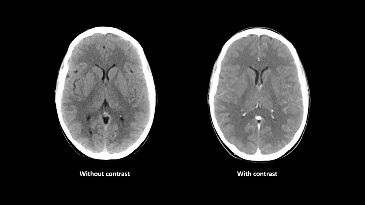
Neuro Imaging
03Thorax, Abdomen, Pelvis Imaging
02Thorax, Abdomen, Pelvis Imaging
01Cardiac Imaging
01Cardiac Imaging
Vascular Imaging
03Pediatric Imaging
Extremities Imaging
01Scan time: 8 s
Scan length: 218 mm
CTDIvol: 6.4 mGy
DLP: 141 mGy*cm
Eff. dose: 0.11 mSv
Courtesy of Luzerner Kantonsspital, Luzern, Switzerland
Without contrast
100 kV, 422 eff. mAs
Scan time: 12.0 s
Scan length: 170 mm
CTDIvol: 40.83 mGy
DLP: 704 mGy*cm
Eff. dose: 1.47 mSv
With contrast
100 kV, 422 eff. mAs
Scan time: 12.0 s
Scan length: 170 mm
CTDIvol: 40.92 mGy
DLP: 706 mGy*cm
Eff. dose: 1.48 mSv
Courtesy of CIMOP Bizet, Paris, France
Scan time: 12 s
Scan length: 49 mm
CTDIvol: 27.08 mGy
DLP: 148 mGy*cm
Eff. dose: 0.46 mSv
Courtesy of CIMOP Bizet, Paris, France
VPCT:
80 kV, 200 mAsÂ
Scan time: 44.0 s
Scan length: 90 mm
CTDIvol: 237.49 mGy
DLP: 2,800 mGy*cmÂ
Eff. dose: 5.88 mSvÂ
°ä°Õ´¡:Ìý
120 kV, 211 mAs
Scan time: 6.0 s
Scan length: 370 mm
CTDIvol: 18.47 mGy
DLP: 724 mGy*cm
Eff. dose: 3.04 mSv
Courtesy of LMU Grosshadern, Munich, Germany
Scan time: 4 sÂ
Scan length: 117 mm
CTDIvol: 3.06 mGy
DLP: 42.6 mGy*cm
Eff. dose: 0.09 mSvÂ
Courtesy of GHI Intercommunal Le Raincy Montfermeil, France
Scan time: 1.9 s
Scan length: 300 mm
CTDIvol: 0.39 mGy
DLP: 15 mGy*cm
Eff. dose: 0.21 mSvÂ
Courtesy of CIMOP Bizet, Paris, France
Scan time: 7.78 s
Scan length: 537 mm
CTDIvol: 17.47 mGy
DLP: 883.56 mGy*cm
Eff. dose: 13.25 mSv
Courtesy of Hopital St. Louis, France
AuSn 120 kV, 467 eff. mAsÂ
Scan time: 13.2 s
Scan length: 468 mm
CTDIvol: 10 mGy
DLP: 486.3 mGy*cm
Eff. dose: 7.3 mSvÂ
Courtesy of University Hospital of Basel, Basel, Switzerland
Scan time: 5.0 s
Scan length: 156 mm
CTDIvol: 8.55 mGy
DLP: 149 mGy*cm
Eff. dose: 1.86 mSv
HR: 61 – 70 bpm
Courtesy of CIMOP Bizet, Paris, France
Scan time: 5.0 s
Scan length: 154 mm
CTDIvol: 13.12 mGy
DLP: 228 mGy*cm
Eff. Dose: 3.19 mSv
HR: 70 bpm
Courtesy of CIMOP Bizet, Paris, France
Scan time: 5.0 s
Scan length: 696 mm
CTDIvol: 2.62 mGy
DLP: 192 mGy*cm
Eff. dose: 2.88 mSv
Courtesy of LMU Grosshadern, Munich, Germany
70 kV, 129 eff. mAsÂ
Scan time: 2.0 s
Scan length: 267 mm
CTDIvol: 1.35 mGy
DLP: 40 mGy*cm
Eff. dose: 0.56 mSv
Courtesy of Linköping University Hospital, Linköping, Sweden
Scan time: 1.1 s
Scan length: 252 mm
CTDIvol: 8.83 mGy
DLP: 260 mGy*cm
Eff. dose: 3.64 mSvÂ
Courtesy of Olmsted Medical Center, Rochester, USA
Scan time: 9.0 s
Scan length: 640 mm
CTDIvol: 2.22 mGy
DLP: 147 mGy*cm
Eff. dose: 2.2 mSv
Courtesy of Linköping University Hospital, Linköping, Sweden
Scan time: 0.6 s
Scan length: 133 mm
CTDIvol: 0.14 mGy
DLP: 2 mGy*cm
Eff. dose: 0.17 mSv
Courtesy of Linköping University Hospital, Linköping, Sweden
Scan time: 8 s
Scan length: 218 mm
CTDIvol: 6.4 mGy
DLP: 141 mGy*cm
Eff. dose: 0.11 mSv
Courtesy of Luzerner Kantonsspital, Luzern, Switzerland
Scanner type | Single Source |
Detectors | Stellar Detector |
X-ray tube | Straton MX |
Max. scan speed | 230 mm/s1 |
In-plane temporal resolution | 142 ms1 |
Rotation time | 0.28 s1 |
kV steps | 70, 80, 100, 120, 140 kV |
mA @ 70 kV, 80 kV, 90 kV | 500 mA @ 70 kV |
Spatial resolution | 0.30 mm |
Table load up to | 307 kg/676 lbs1 |
Gantry opening | 78 cm |
Generator power | 80 kW, 100 kW1 |
Slice acquisition | 128 |
Tin Filters | yes1 |
iMAR | yes1,3 |
As a leading medical imaging company, you can expect exceptional performance, inventiveness, and quality from ecoline, our sustainable and refurbished portfolio of pre-owned systems. ecoline follows externally certified processes, and our very own rigorous 5-step Quality Process ensuring that your system is as good as new – at an affordable price.
The products/features shown on this webpage are not commercially available in all countries. Due to regulatory reasons their future availability cannot be guaranteed. Please contact your local Siemens Healthineers organization for further details.