History
A 59-year-old female patient, suffering from a sudden onset of headache with nausea and vomiting 6 hours previously, was admitted to the hospital. The patient had a medical history of a primary hypertension, stage 3. A CT angiography (CTA) was performed on a conventional energy-integrating detector (EID) CT. This revealed a saccular aneurysm at the C7 segment of the right internal carotid artery (ICA), along with a subarachnoid hemorrhage. An endovascular coiling embolization of the aneurysm was carried out successfully. A follow-up CTA was performed on a dual source photon-counting detector (PCD) CT, NAEOTOM Alpha, for assessment.
Diagnosis
Follow-up CTA images acquired on the PCD-CT showed improved vessel delineation, sharpness and enhanced vessel contrast. However, metal artifacts occurred in the vicinity of the postcoiling aneurysm, obscuring the visualization of the vascular and parenchymal structures. These artifacts were effectively reduced in the images reconstructed with Iterative Metal Artifact Reduction (iMAR) using “Neuro coils” preset, resulting in improved visualization. The aneurysm showed a total occlusion without signs of remnants. The parental right ICA, the anterior cerebral artery (ACA) and the middle cerebral artery (MCA) were all patent without stenosis. The patient recovered uneventfully.
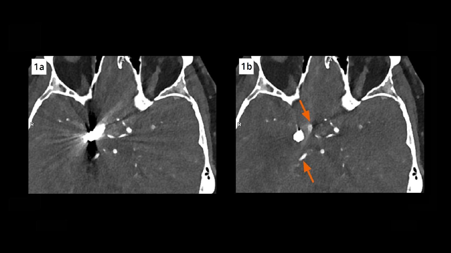
Fig. 1: An axial image prior to iMAR (Fig. 1a) shows an obscured visualization of the embolized aneurysm and the adjacent arteries due to artifacts. The visibility is clearly improved in the image reconstructed with iMAR (Fig. 1b, arrows).
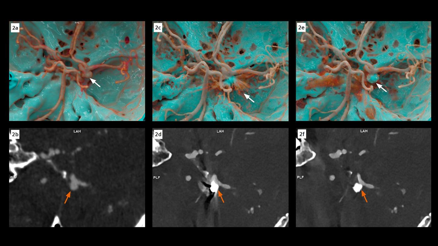
Fig. 2: cVRT and MPR images show the head view of the cerebral arteries. Images acquired with an EID-CT prior to the coiling embolization (Fig. 2a–2b) show an aneurysm located at the C7 segment of the right ICA (arrows). Postcoiling images acquired with PCD-CT without iMAR (Fig. 2c–2d) show metal artifacts obscuring the visibility of the aneurysm and the adjacent structures (arrows). The completely embolized aneurysm, without signs of remnants, as well as patent right ICA, ACA and MCA, are clearly visualized in the images with iMAR (Fig. 2e–2f, arrows). Note the improvement of the vessel delineation, sharpness and contrast in the PCD-CT images (Figs. 2c–2f).
Comments
A ruptured intracranial aneurysm (IA) is an emergency and requires immediate treatment. Endovascular coiling embolization is routinely used to occlude a ruptured IA with the primary aim of preventing the long-term risk of rebleeding. This is associated with a poor prognosis and high morbidity and mortality. [1] Rebleeding can occur either due to a recanalized aneurysm or to a de novo aneurysm, therefore, imaging follow-ups are justified to provide an early preventive treatment when necessary. [2] As MRA is limited by contraindications and DSA is an invasive procedure bearing potential complications such as cerebral thromboembolism, CTA is frequently used for diagnosis and follow-up. However, the challenge lies in the metal artifacts caused by the coils which lead to difficulties in image interpretation, or at worst, non-assessable datasets. To meet this challenge, an iMAR algorithm has been developed which combines beam hardening correction, normalized sinogram inpainting and frequency split to preserve fine anatomical details. The correction process is then iteratively applied for a refined correction result. [3] Consequently, metal artifacts are reduced and the visibility in the vicinity of the postcoiling aneurysm is improved.
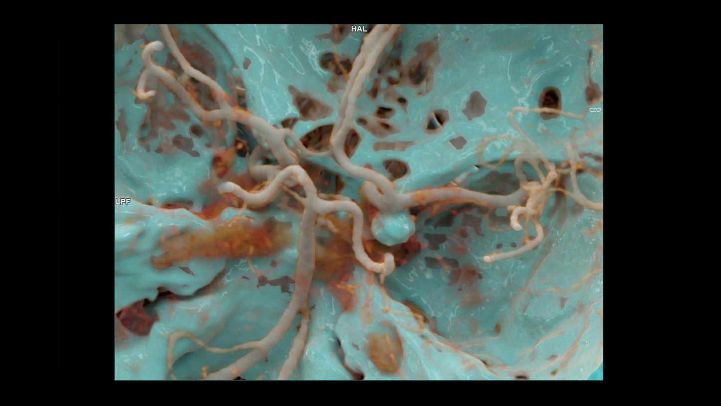
This case is acquired with a PCD-CT, NAEOTOM Alpha, which provides energy-resolved CT data with increased spatial resolution and iodine contrast, without electronic noise. Images are routinely acquired at 0.4 mm slice collimation with spectral information, allowing virtual monoenergetic image (VMI) display at low keV (55 keV in this case), which further enhances image contrast and improves visualization of vascular details. A dedicated sharper kernel (Hv56) is applied in image reconstruction to increase high contrast resolution and improve vessel delineation. These high-resolution images can also be used for three dimensional, in-depth, lifelike demonstration, using cinematic rendering technique (cVRT), facilitating the communication with the neurosurgeons and treatment planning.
As shown in this case, comprehensive PCD CT imaging combined with iMAR technology can improve vessel delineation and sharpness, enhance vessel contrast and reduce metal artifacts. This benefits a follow-up of patients with treated IAs. An interventional procedure for the follow up of this patient was not necessary.
Examination Protocol