Computed Tomography for CardiologyInnovation that matters.
Computed Tomography is a pivotal tool in the battle against cardiovascular diseases, which are responsible for nearly one-third of global deaths.1 CT plays a significant role in addressing the complexities of cardiac cases, from early detection and diagnostic support, to personalized therapy and follow-up.Ěý
The comprehensive suite of one-of-a-kind imaging solutions from Siemens Healthineers deliver the accuracy and speed needed to support cardiologists to make informed, confident decisions.Ěý
As pioneers in cardiac CT solutions, we invite you to witness firsthand how our state-of-the-art CT systems combined with AI-powered technologies are shaping the future of cardiology.
Clinical cases

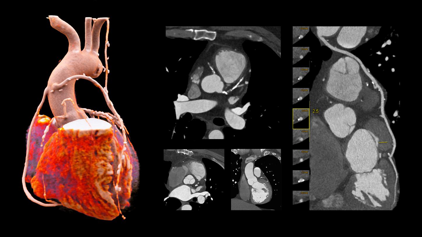
Courtesy of University Medical Center of Freiburg, Freiburg, Germany
NAEOTOM Alpha
Clinical example for challenging conditions – Visualization in the presence of stents, vessel occlusions, and multiple calcifications


Cardiac CT made easy with innovative technologies
White papers
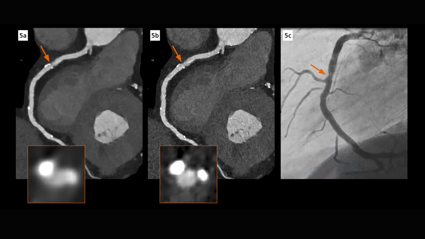
Reach more cardiovascular patients with photon-counting CT
NAEOTOM Alpha with Quantum Technology delivers Quantum HD Cardiac images with 0.2 mm slice thickness. This breakthrough technology not only visualizes previously undetectable details in the heart but does so without dose penalty. Dive deeper into the benefits of photon-counting technology for cardiac CT.
Clinical benefits of Dual Source CT for cardiac imaging
How can you achieve high image quality at minimal radiation dose and contrast media in a coronary CT angiography (CTA) in patients with high heart rates or renal insufficiency?Ěý
Dual Source CT, with its high native temporal resolution of 66 ms, reduces motion artifacts even in high heart rates and also reduces the need for betablockers.Ěý
Download the white paper to explore clinical scenarios where fast native temporal resolution makes a different in cardiac imaging.Ěý
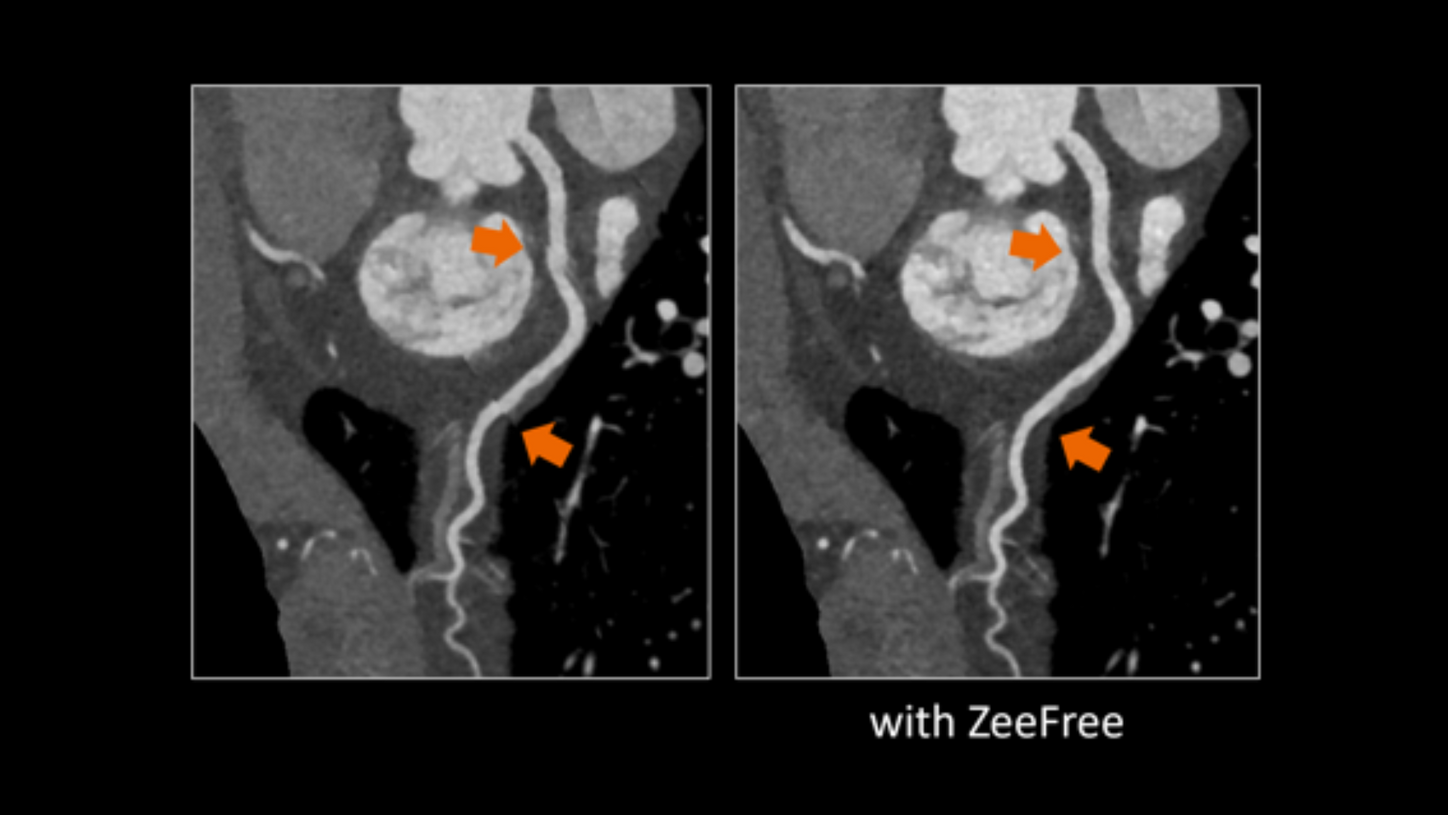
Optimal Cardiac CT imaging with ZeeFree
With ZeeFree, a novel cardiac CT reconstruction feature employing intelligent algorithms to automatically align image stacks, you get images that are optimally aligned to display the morphology as intended, without misalignment of stacks. Zeefree operates on a detector width-independent algorithm, and is adaptable to both retrospective ECG-gated spiral and prospective ECG-triggered sequence acquisitions. Learn more in the white paper.
Customer voices
Ěý