天美影院

Traumatic incudomalleolar dislocation

Kolos Turt贸czki, MD; P谩l Maurovich-Horvat, MD, PhD, MPH; Ibolyka Dud谩s I, MD
Semmelweis University, Medical Imaging Center, Budapest, Hungary

07/23/2025
A 33-year-old female patient, who had sustained injuries following a fall, was presented to the emergency department. Upon arrival, she had altered consciousness with visible bleeding from the right ear and a superficial hematoma on the right side of the skull. A routine head CT scan, as a part of our institutional polytrauma protocol, was performed with a photon-counting detector (PCD) CT scanner (NAEOTOM Alpha.Prime) for further assessment.

CT images showed an incudomalleolar dislocation with a typical 鈥渂roken heart鈥 sign in the thin-slice coronal multiplanar reformats (MPR). The displacement of the head of the malleus from the body and short process of the incus disarticulating the normal 鈥渋ce cream cone鈥 of the joint was also seen in axial images (Fig. 1). These structures were visualized in more details in zoomed reconstructions (Fig. 2). Longitudinal fractures of the right temporal bone (Fig.3a), involving the mastoid cells, the external auditory canal, the middle ear and the ossicles, as well as a complex fracture of the body of the sphenoid bone were present (Fig.3b and 3c). Temporal subdural hematoma, subarachnoid bleeding as well as parenchymal brain contusion injuries on the left, due to the contrecoup mechanism, were seen. There was no evidence of extracranial traumatic lesions. A subsequent control CT examination demonstrated stable contusional injuries with mild progression of the perifocal edema and a regressing subdural hematoma. Immediate neurosurgical intervention was not considered. The patient was treated conservatively on the intensive care unit for two days and was then transferred to the traumatology ward. Ear-nose-throat (ENT) consultation found a right-sided conductive hearing loss.

An axial image shows displacement of the head of the malleus from the body and short process of the incus on the right. A typical 鈥渂roken heart鈥 sign is seen in a coronal MPR image. Two cVRT images created with thin slices demonstrate the incudomalleolar dislocation in three dimensions.
Courtesy of Semmelweis University, Medical Imaging Center, Budapest, Hungary

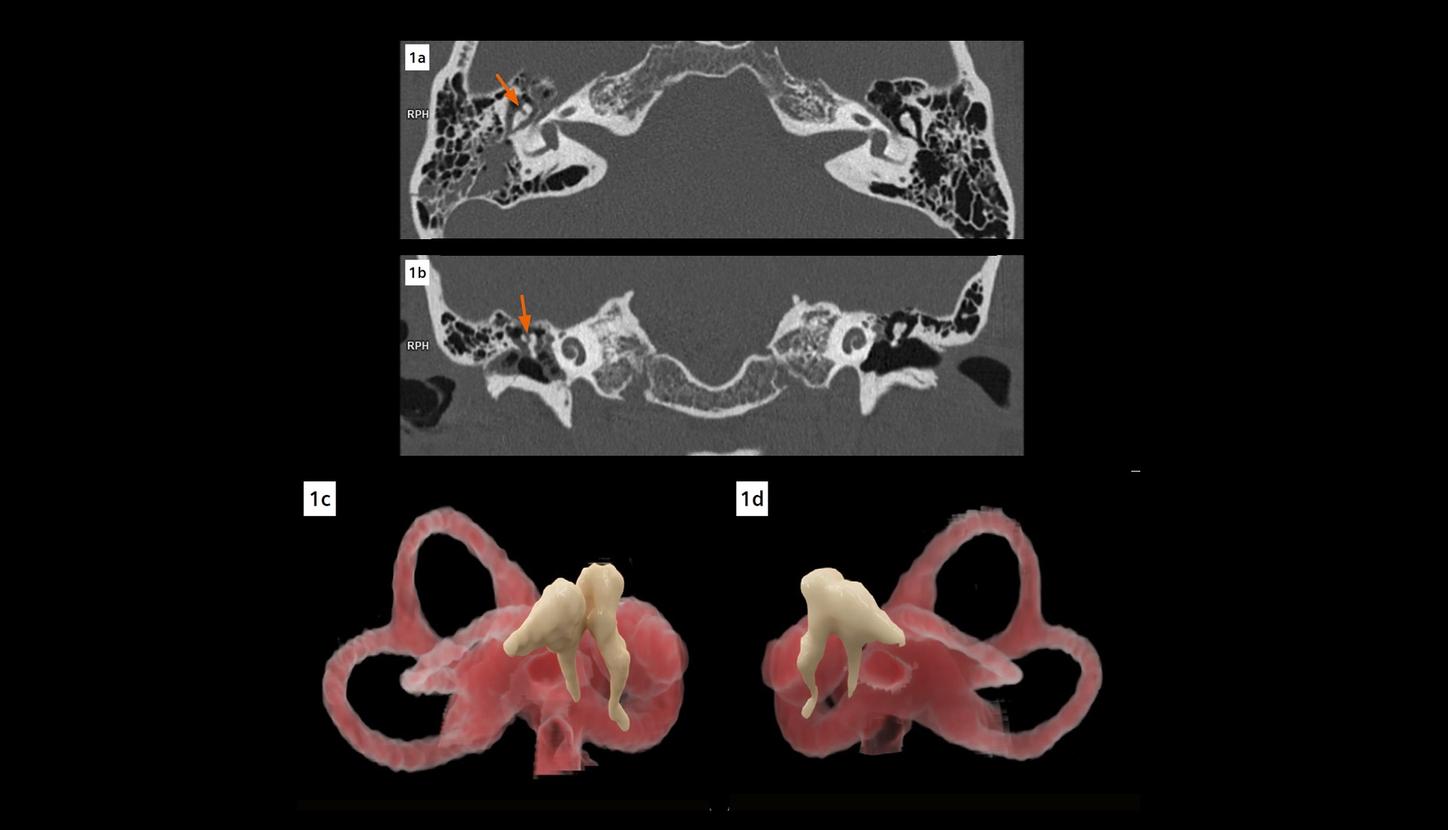
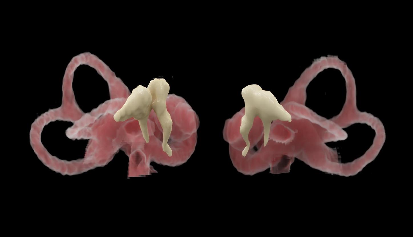
Fig. 1: An axial image (Fig. 1a) shows displacement of the head of the malleus from the body and short process of the incus (arrow) on the right. A typical 鈥渂roken heart鈥 sign (arrow) is seen in a coronal MPR image (Fig. 1b). Two cVRT images (Figs. 1c and 1d) created with thin slices (0.4 mm, sharp kernel of Hr76) demonstrate the incudomalleolar dislocation (Fig. 1c) in three dimensions.

Images from zoomed reconstructions show the incudomalleolar dislocation with signs of disarticulated 鈥渋ce cream cone鈥 and 鈥渂roken heart鈥 in details.
Courtesy of Semmelweis University, Medical Imaging Center, Budapest, Hungary

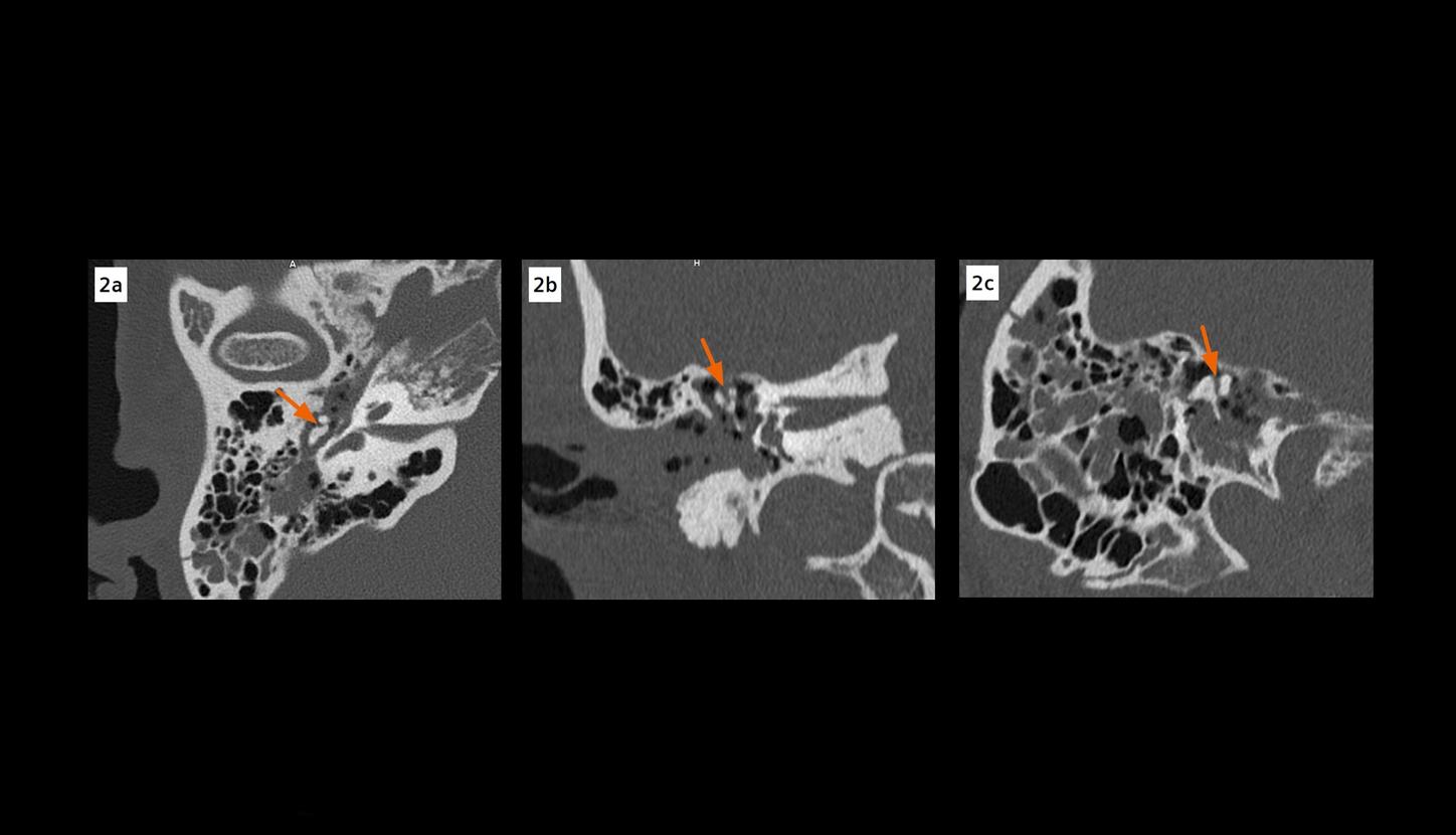
Fig. 2: Images from zoomed reconstructions show the incudomalleolar dislocation (arrows) with signs of disarticulated 鈥渋ce cream cone鈥 (Fig. 2a) and 鈥渂roken heart鈥 (Fig. 2b) in details.

cVRT images show longitudinal fractures of the right temporal bone, involving the mastoid cells and the external auditory canal, as well as a complex fracture of the body of the sphenoid bone.
Courtesy of Semmelweis University, Medical Imaging Center, Budapest, Hungary

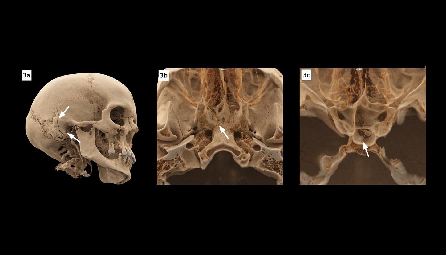
Fig. 3: cVRT images show longitudinal fractures (Fig. 3a, arrows) of the right temporal bone, involving the mastoid cells and the external auditory canal, as well as a complex fracture of the body of the sphenoid bone (Fig. 3b and 3c, arrows).

Temporal bone trauma is commonly encountered in the emergency department. The tympanic cavity and tegmen, the ossicular chain, the bony labyrinth, the facial canal, the internal carotid artery, the jugular foramen and venous sinuses, as well as the intracranial contents are all potential injury sites. A specific and thorough reporting approach is needed due to the unique composition of these anatomical structures. A delayed or missed diagnosis of injuries to these structures can result in severe and permanent disabilities for the patient. [1] Ossicular luxation or fracture, disrupting the conductive chain, may cause conductive hearing loss (CHL). Six months after temporal bone trauma, persistent CHL occurs in 50% of the patients. The most common sites of ossicular dislocation are the incudostapedial and the incudomalleolar joints. [2] In the latter case, the joint dislocation is marked with displacement of the scoop (head of the malleus) from the cone (body and short process of the incus), disarticulating the normal 鈥渋ce cream cone鈥 in axial images and the typical 鈥渂roken heart鈥 sign in coronal MPR. [3] [4] CT is the imaging modality of choice in cases of temporal bone trauma. Traditionally, CT examination for head and for temporal bones are two separate scans, with the former focused on the low-contrast cerebrum and the latter, on high-contrast bony structures. This case is acquired with a newly developed single source PCD CT, NAEOTOM Alpha.Prime. The principle of PCD differs from that of conventional, energy-integrating detector (EID). With EID, X-ray photons are first converted into scintillation light and then into electrical signals, while with PCD, X-ray photons are converted directly into electrical signals. The detector elements in EID are separated by physical separators, while in PCD, they are separated by electric fields. The electronic circuit noise is also eliminated in PCD. These differences result in increased efficiency of X-ray photon utilization, and potentially, reduced image noise and radiation dose. Additionally, PCD has a smaller detector pixel size, which contributes to the decreased partial volume effects and blooming artifacts. This increases spatial resolution, which is especially important for imaging small, high-contrast structures, such as the ossicular chain. [5] Images acquired in routine head scan can be reconstructed both at 0.4 mm slice width with a sharp kernel (Hr76) for visualizing the temporal bones and at thicker slice width with a standard or smooth kernel for cerebrum imaging, obviating the necessity of having to do two separate scans. These high-resolution images can also be used to create three dimensional images, using cinematic rendering technique (cVRT), providing detailed, lifelike visualization. In trauma settings, PCD CT, with its increased spatial resolution in routine scans, may enable radiologists to reach diagnostic conclusions that were previously only possible with high resolution CT (HRCT) examinations.

Scanner

NAEOTOM Alpha.Prime

Scan area

Head

Scan mode

Quantum

Scan length

182 mm

Scan direction

Cranio-caudal

Scan time

69 s

Tube voltage

120 kV

Effective mAs

167 mAs

IQ level

225

Dose modulation

CARE Dose4D

CTDIvol

28 mGy

DLP

554 mGy*cm

Rotation time

0.5 s

Pitch

0.35

Slice collimation

144 x 0.4 mm

Slice width

0.4 mm

Reconstruction increment

0.2 mm

Reconstruction kernel

Hr76

Subscription Button Icon
Be the first to know about our events, training, and news