
Over a quarter century ago, PET/CT was first introduced to the medical community. After an initial tepid reception, the newly combined modality was enthusiastically integrated into medical practice and is now considered to be an indispensable tool. Two pioneers of PET/CT share their first impressions of what it was like to be an early adopter and apply the modality into their clinical practice and research—and, ultimately, what it means to patients.
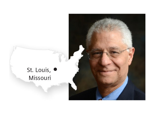
Barry A. Siegel, MD
Mallinckrodt Institute of Radiology at Washington University School of MedicineĚýand Barnes-Jewish Hospital, St. Louis, Missouri, USA
After receiving his medical degree from Washington University School of Medicine in St. Louis, Missouri, Barry A. Siegel, MD, remained at Washington University for his residency training. He became a professor of radiology and medicine and director of the Division of Nuclear Medicine at the university and at the affiliated Barnes-Jewish Hospital. In 2021, the American College of Radiology’s Gold Medal Award recognized Siegel for his more than four decades of Leadership within the nuclear medicine community. This award was bestowed for helping to develop PET as an important clinical and research tool and advancing the quality of imaging clinical trials.
Ěý
Tell us about your first experience with PET/CT.
We got our first PET/CT (Biograph Duo) in 2001 at Barnes-Jewish Hospital. We were immediately impressed, as were patients, at the shortened exam time (cutting the transmission imaging from nearly 20 minutes for a body study to less than a minute). This was a boon for both us and for patients. Anatomic correlations became essentially painless. With our ECAT scanner, we had a monitor for reviewing the PET images on one counter and behind us were the CT images on film on a view box. Pretty quickly we realized what an improvement the fused PET/CT was. In those early days, the image review software was a bit clunky, but we and others quickly realized what changes were needed, and Siemens and other vendors made this happen. Most of our studies were body studies for Cancer indications, such as pulmonary nodule evaluations, initial staging of non-small cell lung cancer, and detection of suspected recurrent colorectal cancer, and lymphoma, since those were the only ones that were then covered by Medicare (and other insurers closely followed what Medicare did). And then we started the National Oncologic PET Registry (NOPR) which eventually led to broader coverage.1
Describe a significant moment in the past 25 years with PET/CT.
One of my favorite recollections (however not a blockbuster) was related to fludeoxyglucose injection F 18 (FDG) uptake in brown fat. I remember a number of luminaries presenting PET-only images at meetings and saying that uptake was in lymph nodes (especially when in the upper mediastinum) or in muscle (which was a theory I often espoused), and then the papers from Zurich University (Zurich, Switzerland) and Johns Hopkins University (Baltimore, MD, USA) came out and proved how foolish we were.
Please share your thoughts on PET/CT’s clinical role in Patient care today.
It is inestimable. PET/CT is a central component of oncology practice. Patient management would be greatly impaired without it. PSMA-PET has been transformative for evaluating men with prostate cancer. The role in central nervous system (CNS) imaging, a rapidly growing area, is central to selection of therapy for Alzheimer’s disease. Cardiac imaging has come along the slowest, but I see a day where the majority of myocardial perfusion imaging (MPI) studies are performed with PET/CT.

Richard Wahl, MD
Mallinckrodt Institute of Radiology at Washington University School of Medicine and Barnes-Jewish Hospital, St. Louis, Missouri, USAĚý
Richard L. Wahl, MD, received his medical degree from Washington University School of Medicine in St. Louis, and later became director of the Division of Nuclear Medicine at Johns Hopkins University. He is currently a professor at Mallinckrodt Institute of Radiology (MIR) at Washington University School of Medicine in St. Louis. He served as the director of MIR from 2014 to 2023. In 2018, Wahl was bestowed the Georg Charles de Hevesy Award from the Society of Nuclear Medicine and Molecular Imaging, where he served as president in 2021-2022.
Tell us about your first experience with PET/CT.
I was an early adopter of FDG PET and recall using our early whole-body PET scanner at the University of Michigan (Ann Arbor, MI, USA) to image patients with breast cancer, melanoma, lymphoma, and lung cancer. The target to background ratios were high and we had “hot spots” on PET but were guessing as to where they were on CT.2 While we used the transmission images as crude localizers in the chest, they were useless in the abdomen and pelvis. So, we developed software fusion methods to generate PET fused with CT and PET fused with MRI images, which we called “anatometabolic images.”3
We showed that the FDG-PET-fused-with-CT images were more accurate than a CT scan for staging NSCL cancer. This was a major accomplishment and paved the way for the routine use of FDG-PET in this setting.
But the software fusion took forever and did not always work. A whole new world opened when we installed our first PET/CT at Johns Hopkins in the spring of 2001. What took us a week with the software was done in 30 minutes with the PET/CT. It sped up patient throughput and consistently improved diagnostic accuracy versus PET alone. It was “immediately obvious” this was the future of PET.
Describe a significant moment in the past 25 years with PET/CT.
The early days were remarkable seeing new things every day. Figuring out that brown fat was FDG-avid and widely distributed and that many bone metastases were “invisible” on CT but seen on PET/CT were early highlights and somewhat unexpected. I also learned how many things I had missed detecting on CT alone and got better at CT because of PET/CT.
Please share your thoughts on PET/CT’s clinical role in patient care today.
PET/CT is indispensable in the management of many cancers and a true workhorse in our clinic. It has been very exciting to see Technology disseminated across the world to the benefit of millions of patients each year. I am delighted to have access to high-throughput, high-sensitivity PET scanners for my clinical work.
Fludeoxyglucose F 18
Please see Indications and Important Safety Information for Fludeoxyglucose F 18 (18F FDG) Injection.