
SOMATOM X.cite eco
with myExam CompanionIntelligent imaging. Excellence empowered.
ecoline - Refurbished. Sustainable. As good as new.
The number and complexity of radiological procedures are increasing. This calls for new solutions: Intelligent navigation. Patient-friendly design. Personalized imaging. Consistent standards.
SOMATOM® X.cite offers all that. Together with the unique guidance of myExam Companion and myNeedle Companion, the CT scanner enables excellent clinical decisions in diagnostic and interventional procedures. Find out how SOMATOM X.cite empowers excellence in computed tomography – from routine to advanced procedures.
Features & Benefits

SOMATOM X.cite with myExam Companion facilitates scans and workflows
Intelligent navigation for enhanced consistency
With its two digital Companions, SOMATOM X.cite provides unique guidance to support you in your daily work. myExam Companion offers intuitive, interactive guidance through any CT scan. myNeedle Companion helps simplify targeted needle placement even in complex CT interventions. And our Mobile Workflow allows you to spend more time at your patient’s side.
Intelligent cardiac imaging in CT
Find out how SOMATOM X.cite with myExam Companion supports navigating any cardiac CT like a routine exam.
Intelligent neuro imaging in CT
Discover how you can acquire fast and consistent CT results with SOMATOM X.cite with myExam Companion.
Intelligent spectral imaging in CT
Learn how you can easily gain additional functional information in CT imaging with SOMATOM X.cite with myExam Companion.

Mobile Workflow: A whole new way to operate the scanner
Stay close to your patients: Prepare a scan via tablet, start it with the remote control, keep an eye on patients even when they are inside the gantry, and preview the images directly on the tablet.
Customer voices


Extra room to help patients feel relaxed

Patient-friendly design with an 82 cm bore
From enhanced interfaces that improve communication between you and your patient to the large bore and the tablet-based Mobile Workflow: SOMATOM X.cite is designed to transform how patients interact with both you and the CT scanner and how they perceive their care.
Customer voices

Drive standardization across modalities
Consistent standards across your institution
myExam Companion, myNeedle Companion, and Shui® – the design system of Siemens Healthineers – provide you with common interfaces across multiple modalities. Combined with our digital solutions, they can help you redefine and standardize protocols across your institution, optimizing clinical workflow, staffing schedules, results, and productivity.

Automated guidance
myExam Companion helps optimize scan parameters individually for every patient – and supports consistent image quality across your fleet.

One user interface across modalities
Clinical Use
Personalized imaging for improved diagnostic confidence
SOMATOM X.cite generates the comprehensive information that can help you diagnose with precision and confidence – no matter the patient or procedure. The power stems from its outstanding imaging chain, which includes the Vectron® X-ray tube. The user guidance comes from myExam Companion, which tailors acquisition to the individual patient.
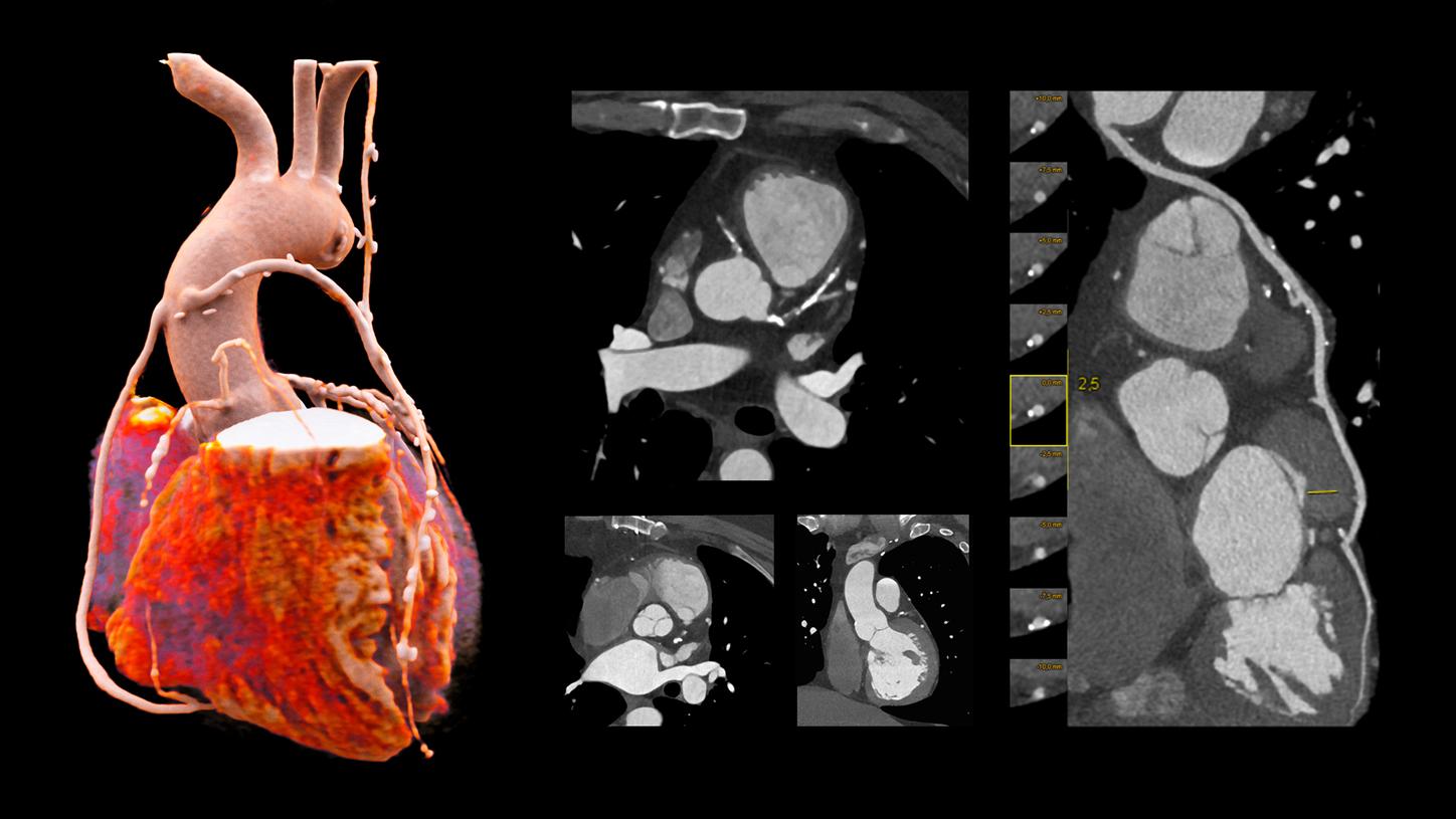
Adaptive Cardio Spiral
120 kV
CTDIvol:Ěý13.8 mGy
DLP: 302.2 mGy*cm
Exposure time: 10 s
Scan length: 219 mm
Rotation time: 0.3 s
HR: approx. 50 bpmĚý
- Robust cardiac imaging with myExam Companion
- Enhanced delineation of soft and hard plaque thanks to ADMIRE iterative reconstruction
- Cinematic VRT
- 0.8 mm MPRs used for reconstructions
- Curved MPR of LAD
Courtesy of University Hospital Erlangen, Erlangen, Germany


Technical Specifications
ecoline - Refurbished. Sustainable. As good as new.
As a leading medical imaging company, you can expect exceptional performance, inventiveness, and quality from ecoline, our sustainable and refurbished portfolio of pre-owned systems. ecoline follows externally certified processes, and our very own rigorous 5-step Quality Process ensuring that your system is as good as new – at an affordable price.
Did this information help you?
The statements by Siemens Healthineers’ customers described herein are based on results that were achieved in the customer’s unique setting. Since there is no “typical” hospital and many variables exist (e.g., hospital size, case mix, level of IT adoption) there can be no guarantee that other customers will achieve the same results.
Tested and validated for DICOM Images from CT scanners of Siemens Healthineers, GE Healthcare, and Philips Healthcare.