A 47-year-old male presented with persistent upper abdominal pain. A conventional CT scan found an ill-defined hypo-enhancing mass in the pancreatic body, suspicion of intraductal tumor spread, and a potential liver metastasis. With informed consent, another CT scan was performed using an ultra-high resolution mode on a photon-counting CT, NAEOTOM Alpha.
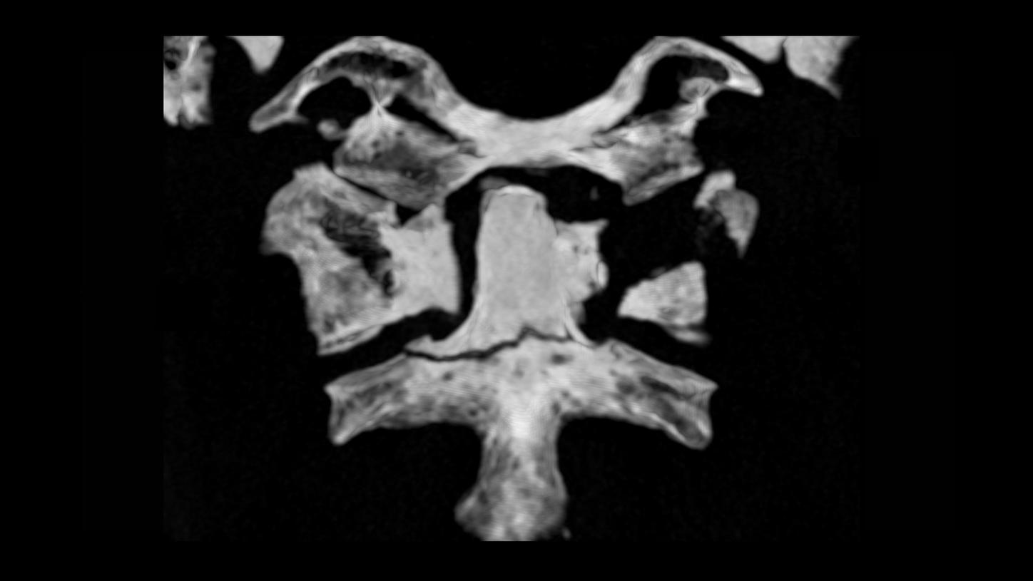
A 65-year-old male patient, struck by a car while cycling, was admitted to the Department of Traumatology. A CT scan was performed on an energy-integrating detector CT, which revealed multiple fractures of the cervical spine. 24 hours later, a control CT examination was performed on a single source photon-counting detector CT, NAEOTOM Alpha.Prime.
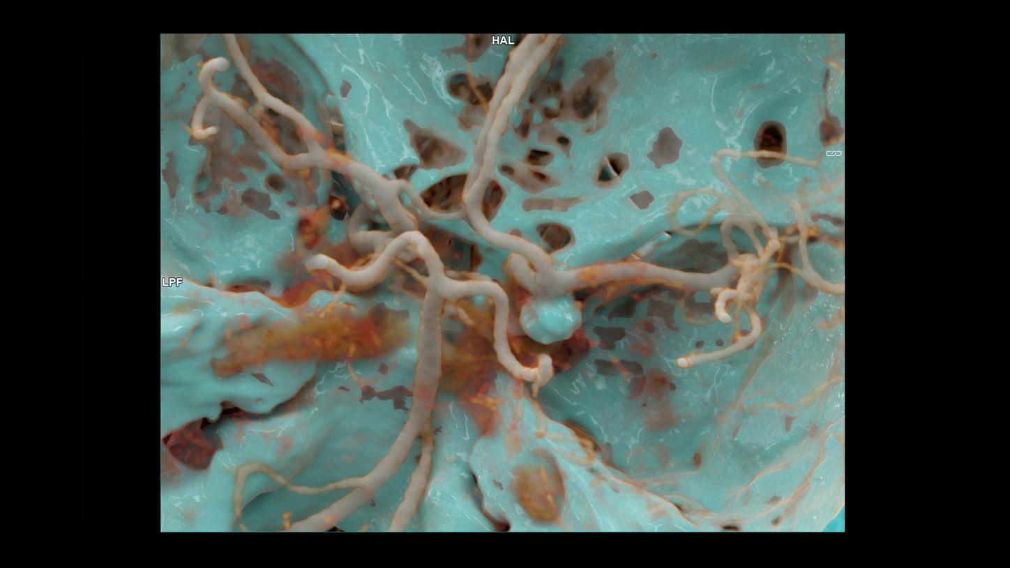
A 59-year-old female patient, suffering from a sudden onset of headache with nausea and vomiting, was diagnosed with a saccular aneurysm at the internal carotid artery and subarachnoid hemorrhage. After a successful endovascular coiling embolization of the aneurysm, a follow-up CT angiography was performed on a dual source photon-counting CT, NAEOTOM Alpha.
A 15-year-old male patient, complaining of a growing lump on the right of his forehead over the past 20 days, came to the hospital for a check-up. A contrast-enhanced CT scan was performed with a dual source photon-counting CT, NAEOTOM Alpha, using an ultra-high resolution scan mode, for assessment.
A 47-year-old female patient, complaining of effort dyspnea, came to the hospital for a check-up. A performed echocardiography showed a slightly dilated left ventricle with a mid-range ejection fraction and systolic function. A cardiac CT angiography was performed on a photon-counting CT, NAEOTOM Alpha, to assess the coronary arteries.
A 5-month-old baby, suffering from severe hypertension, was presented to the hospital. Ultrasound examination showed bilateral congenital hydronephrosis. A contrast CT scan was performed with a dual source photon-counting detector CT, NAEOTOM Alpha.Pro, to rule out renovascular etiology of the hypertension prior to pyeloplasty.
A 2-year-old dog was rescued after being abandoned at a lodge. An abdominal ultrasound examination suggested a possible portosystemic shunt. Surgery was attempted, however, failed to find the anomaly. An abdominal CTA was requested to identify the suspected anomalous vessel.
A 71-year-old female patient, complaining of dizziness for over two months, came to the hospital for a check-up. A native cerebral CT and a CT angiography (CTA) were performed on an energy-integrating detector (EID) CT for assessment.
A full-term neonate was born with a suspicious congenital heart malformation. Prior to the planned surgical repair, a cardiac CT examination was requested to evaluate the heart anatomy, the great arteries and the coronary arteries.
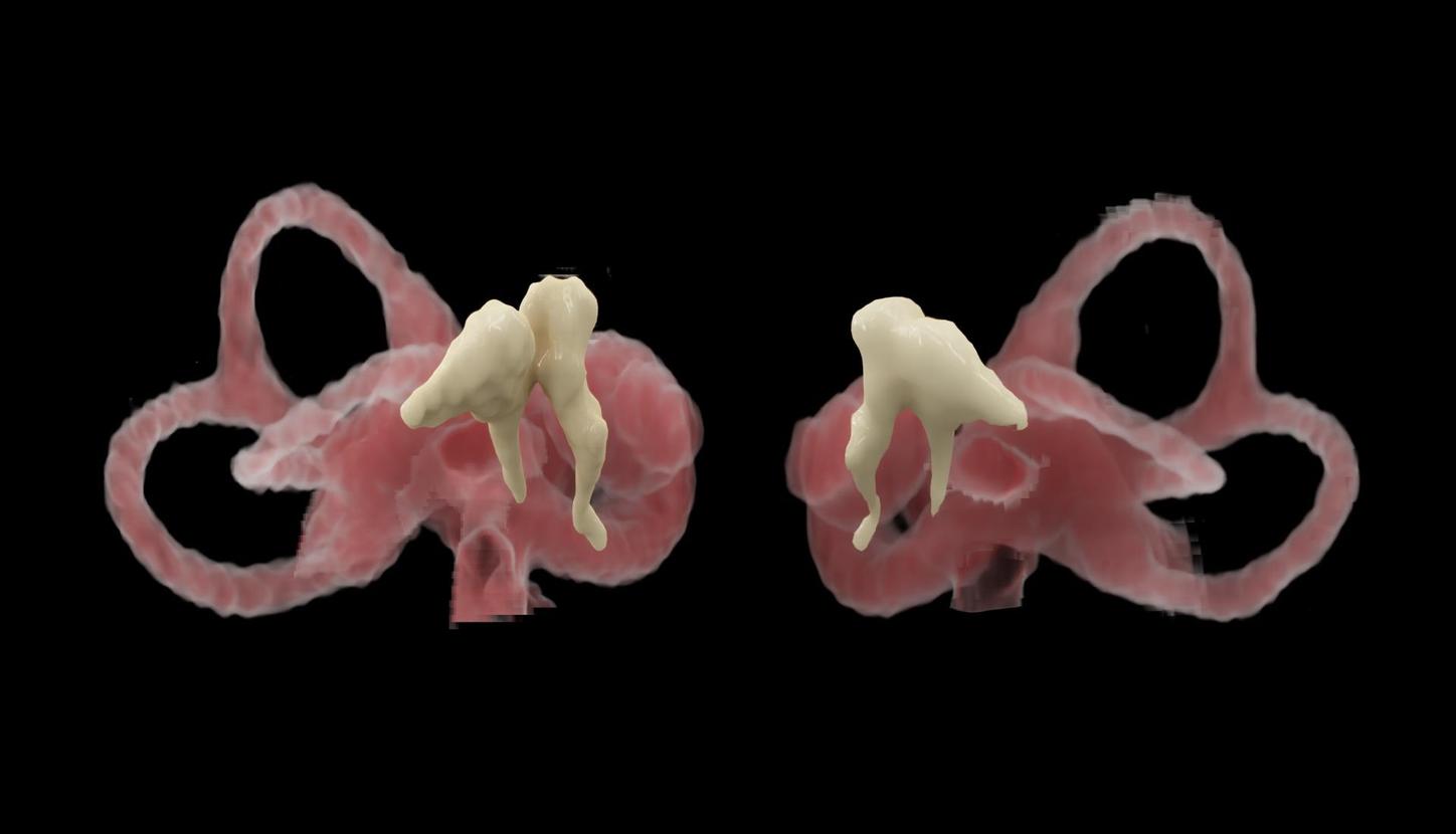
A 75-year-old female patient was presented to the hospital due to an episode of instability. A routine examination revealed a progressive conductive hearing loss on the right side. An ultra-high resolution scan was performed using a dual source photon-counting detector CT, NAEOTOM Alpha®, to investigate the etiology of her hearing loss.
A 53-year-old female patient, complaining of abdominal wall varices, progressive abdominal tenderness and fatigue for the past two years, came to the hospital for a check-up. A triple-phase (arterial, portal-venous and delayed) contrast-enhanced abdominal CT examination was requested and performed for assessment.