


Press release
Siemens Healthineers Expands Contrast-Enhanced Mammography and Biopsy Capabilities of Mammomat B.brilliant
Booth #2529 RSNA 2025, Chicago, IL
Published on November 30, 2025
Image Gallery
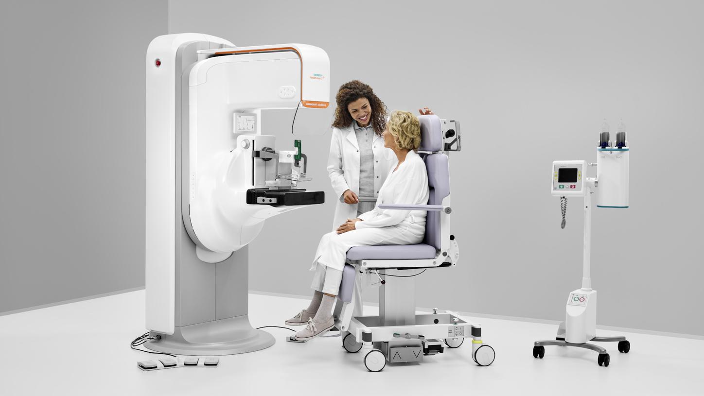
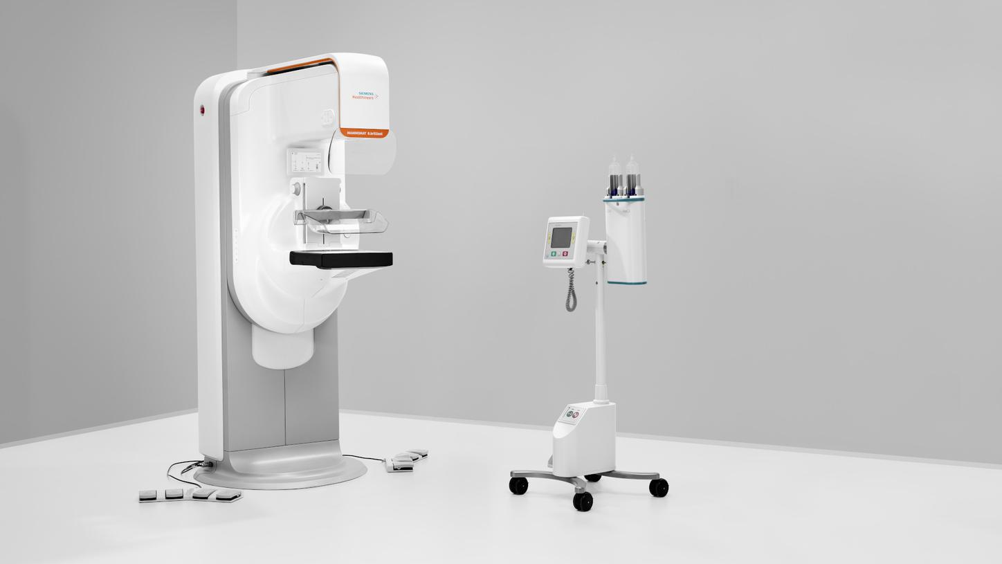
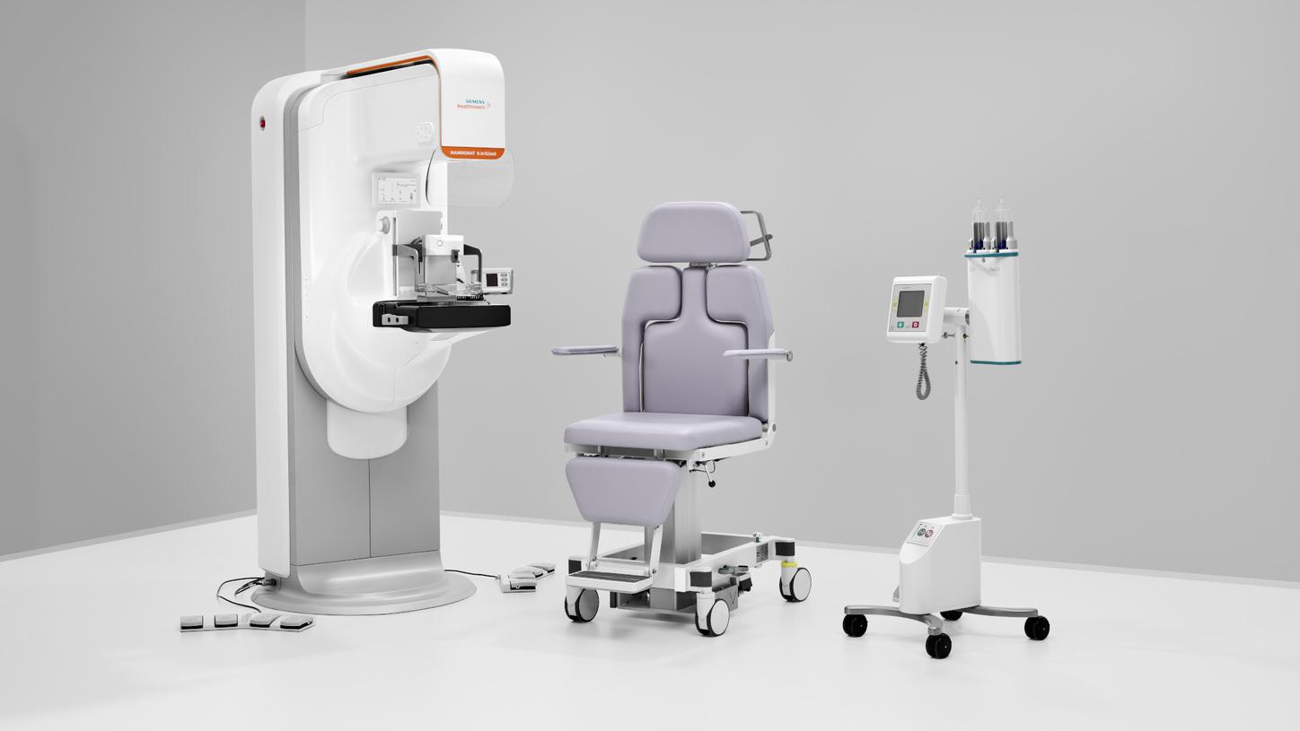
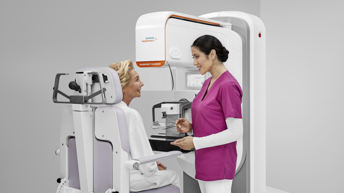
ClearCEM technology on Mammomat B.brilliant2 delivers outstanding image quality, ushering in a new era of contrast enhanced mammography.
Source: Siemens Healthineers
(pdf, 186.01 KB)
Contact

Stefanie Haug
Magnetic Resonance Imaging, Computed Tomography and X-ray
1Data on file. For average breast size of 50/50 glandular/adipose tissue and 5 cm thickness.
2ClearCEM with Mammomat B.brilliant VA11 is pending 510(k) clearance, and is not yet commercially available in the USA. Mammomat B.brilliant is not commercially available in all countries. Due to regulatory reasons its future availability cannot be guaranteed.
3The statements by customers of Siemens Healthineers described herein are based on results that were achieved in the customer's unique setting. Because there is no โtypicalโ hospital or laboratory and many variables exist (e.g., hospital size, samples mix, case mix, level of IT and/or automation adoption) there can be no guarantee that other customers will achieve the same results. Dr. Diane Georgian-Smith receives financial support from Siemens Healthineers for collaborations.
Siemens Healthineers 2026
Siemens Healthineers pioneers breakthroughs in healthcare. For everyone. Everywhere. Sustainably. The company is a global provider of healthcare equipment, solutions and services, with activities in more than 180 countries and direct representation in more than 70. The group comprises Siemens Healthineers AG, listed as SHL in Frankfurt, Germany, and its subsidiaries. As a leading medical technology company, Siemens Healthineers is committed to improving access to healthcare for underserved communities worldwide and is striving to overcome the most threatening diseases. The company is principally active in the areas of imaging, diagnostics, cancer care and minimally invasive therapies, augmented by digital technology and artificial intelligence. In fiscal 2025, which ended on September 30, 2025, Siemens Healthineers had approximately 74,000 employees worldwide and generated revenue of around โฌ23.4 billion. Further information is available at siemens-healthineers.com.